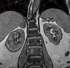
MRI Adrenal
An adrenal gland is an organ that is situated on top of your kidneys and produces hormones.
Preparation
You will need to arrive 30 minutes before your appointment time to allow preparation for your examination Bring any previous imaging with you that is relevant to the area we are scanning, such as x-rays, CT scan and any previous MRI's you may have had. All patients (except for diabetics) are required to fast for four (4) hours prior to your appointment time. You may drink clear fluids but do not eat. As the MRI uses a very strong magnet, no metallic objects or mechanical devices can enter the imaging room. You may want to keep this in mind when deciding what to wear to your appointment. Below is a list of suggestion to help you prepare for your appointment:
You will be asked to complete an MRI Patient screening form prior to you exam. (link to MRI Screening safety form) Additional information or testing may be needed prior to your MRI scan to ensure that it is safe for you to have this test, especially:
Procedure
You will need to remove any items of clothing with metallic objects such as belts, bras and zippers and a hospital gown will be given for you to wear if required. You will be allocated a locker for your personal belongings to be kept safe during the MRI scan. During the MRI exam, you will be lying on a firm table. A piece of equipment called a coil will be placed over your abdomen. This device is essential for the sending and receiving of radio waves that produce the image. The radiographer will then move the table to the centre of the MRI machine. The inside of the machine is like a giant tunnel that is well lit and open at each end. You will need to lie completely still for the duration of the scan so ensure you are comfortable before it begins. The MRI makes loud knocking noises while we take the image and we will provide you with ear plugs to reduce the noise. The radiographer will leave the room on the commencement of the scan but will be able to see you through a window. There are speakers located inside the MRI to enable you and the radiographer to communicate if you need to. You will also be given a call bell if there are any problems during the procedure. You will have a needle placed in your arm and an injection for an injection of the MRI contrast, called gadolinium to be given. Results
A specialised doctor will carefully analyse your images and make a report for your referring doctor. If the referring doctor is in the hospital they will be able to access the results on their computer. If you are an out-patient and require a copy of the images then a disc of images can be made available within 5 working days after the MRI scan.
|

